Portrait of Dr. Asif Mehmood
Computational neuroscience · neuromorphic AI · biomedical engineering
PhD Computer Science · UET Taxila · 2025

Dr. Asif Mehmood

I build spiking models of excitable cells (neurons, cardiac myocytes and pancreatic cells) and use them to study how the brain and heart talk to each other, to detect disease early from electrophysiological signals, and to simulate networks at human-brain scale on a desktop computer. My next step takes these models from simulation to the patient: biosensors, embedded signal processing and mobile apps that flag anomalies in the brain, heart and pancreas.

86%coincidence factor on 2,191 recorded neurons
83%firing-rate prediction accuracy
1011neurons simulated in about 13 GB
5papers, first author on all
CH1 · neuron VN CH2 · cardiac myocyte VC
Tonic-spiking neuron + human ventricular action potential · press Run live2 s / sweep · 50 mV / div
Research themes

One cell model, four lines of work

My doctoral work proposed the Hybrid Excitable Cell (HEC) model: a computationally light spiking model with an adaptive threshold, neurotransmitter dynamics and adrenergic receptors. Everything else on this page grows from that model, and it sits where neuroscience, cardiology, endocrinology, electronics and AI meet.

Unified excitable cells

A hybrid model for neurons, heart and pancreas

A single set of equations that reproduces 14 neurocomputational firing patterns, cardiac action potentials of eight species, and pancreatic cell activity. The adaptive threshold carries spike-timing history, and adrenergic receptors give the bidirectional brain–organ link.

HEC model membrane potential with adaptive threshold
Brain–heart interaction

Cardio-neural networks and heart rate variability

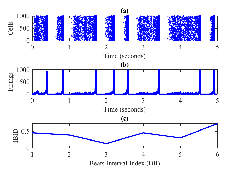
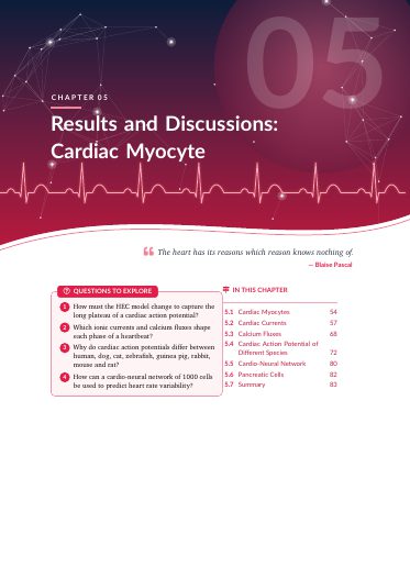
A fully connected network of 1,000 neurons and cardiac myocytes, linked through α- and β-adrenergic neurons, reproduces the heartbeat as collective neural firing. Heart rate variability (RMSSD) is read directly from the simulated electrophysiology.

Cardio-neural network raster, beats and inter-beat differences
Brain-scale simulation

100 billion neurons on a desktop CPU

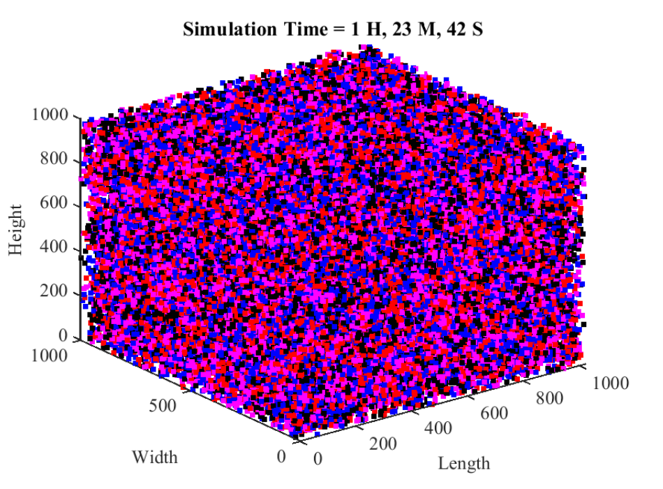
Storing each neuron's response as one bit, packed 64 to a word, brings a human-brain-size network of 1011 neurons and 1014 synapses down to about 13 GB. The full run took 1 h 23 min on a Core i5 laptop with 4 GB of RAM.

3-D firing map of the 100-billion-neuron simulation
Spiking AI for healthcare

Anomaly detection in brain, heart and pancreas

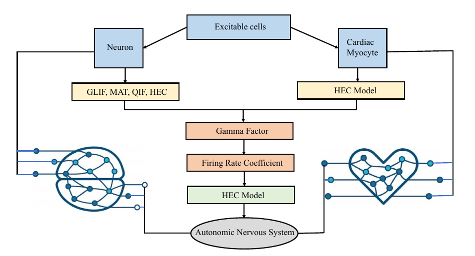
Hybrid spiking neural networks built on HEC neurons flag abnormal patterns in neural, cardiac and pancreatic signals and predict cardiovascular disease, with the energy profile that suits wearable and implantable devices.

Flowchart of the brain–heart communication pathway
Research directions

From cell model to patient: a sensor-to-smartphone pipeline

The next stage of my work closes the loop between the models and real people. Biosensors capture the brain, heart and glucose signals; embedded electronics clean and digitise them; HEC-based spiking networks read them; and an Android app warns the user and their clinician when something looks wrong. My background in electronics (MSc Electronics, 15+ years teaching electrical and electronics engineering) is what makes this hardware-to-AI chain possible.

Stage 1 · Sense

Sensor interfacing & biosensor integration

Wearable and implantable sensors for the three organs, connected to one acquisition front end.

  • EEG electrodes, ECG / PPG patches
  • Electrochemical glucose biosensors (CGM)
  • Analogue front ends, ADCs, low-power BLE links
Stage 2 · Condition

Electronic & biomedical signal processing

Real-time filtering and feature extraction on embedded hardware, close to the sensor.

  • Artefact and noise removal, adaptive filtering
  • R-peak / HRV, EEG rhythm and glucose-trend features
  • Microcontroller, DSP and FPGA implementation
Stage 3 · Understand

Neuromorphic & edge AI

HEC spiking networks and physiological digital twins that learn each patient's normal pattern.

  • Event-driven, energy-efficient inference
  • Anomaly detection with explainable outputs
  • Brain–heart–pancreas coupling models
Stage 4 · Act

Android apps for anomaly detection

A mobile-health app that shows the signals, raises alerts and shares reports with clinicians.

  • On-device models (TensorFlow Lite / ONNX)
  • Seizure, arrhythmia and hypo/hyperglycaemia alerts
  • Remote monitoring and telemedicine
Computational neuroscienceNeuromorphic engineeringBiomedical engineeringCardiology & HRVDiabetes & endocrinologyEmbedded systems & IoTDigital health / mHealthBrain–computer interfacesAI for healthcareHigh-performance computing
Simulation 1 · Neuron lab

Fire a model neuron in fourteen different ways

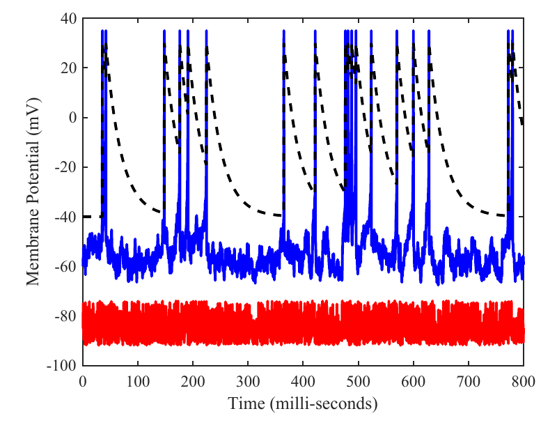
Pick a firing pattern from Table 3.1 of my thesis, change the stimulus and add correlated noise. The trace is drawn the way the thesis plots it: membrane potential in blue, adaptive threshold dashed, input current in red.

Firing pattern
τv′ = k(v−Vr)f(v−Vth) − u + RI  (3.1)
τu′ = a(bv − u)  (3.2)
Vth = A·e(ts−t)/τ − θ  (3.5)
Regular spiking
membrane potential v (mV)adaptive threshold Vthinput current I (offset)
0spikes
0mean rate (Hz)
–first-spike latency (ms)
–mean ISI (ms)

In-browser demo, forward Euler integration as in Algorithm 1. It uses the quadratic Izhikevich-form dynamics and published presets from which the Table 3.1 parameters were tuned; the dashed line plots the adaptive threshold of Eq. 3.5 after each spike.

Results

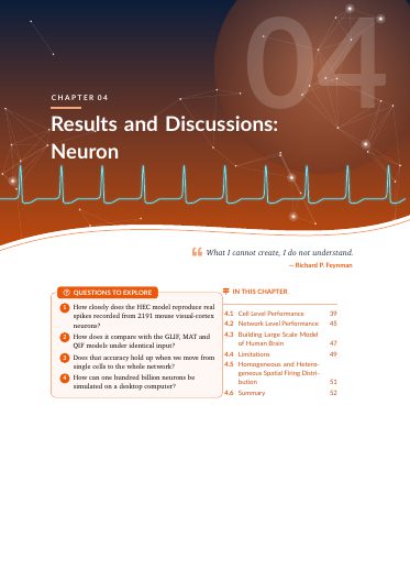
Tested against 2,191 real neurons

Electrophysiological recordings of 2,191 neurons from mouse visual cortex, stimulated optogenetically, over 14 trials of 217 time bins. The HEC model beats GLIF, MAT and QIF on spike-timing and network firing rate.

Coincidence factor, trial 1 (Table 4.1)

spikes coinciding with the recording

Computational cost

FLOPs per update · log scale (Fig. 4.4)

Γ across 14 trials (Table 4.3)

hover for values
Show as table
Simulation 2 · Brain–heart

Watch 1,000 neurons keep time with a heart

Each cardiac upstroke fires β-adrenergic neurons; each repolarisation fires α-adrenergic neurons. Their summed firing marks the heartbeat, and the beat-to-beat intervals give heart rate variability, as in Chapter 5 and the Neuroinformatics paper.

Neuron parameters follow §5.5: a, b, c = −65 + 15r², d = 8 − 6r², with r uniform on [0, 1].

Cardiac myocyte action potentiallast 3 s
● β-neurons  ● α-neurons · 1,000 cellslast 5 s
Population firing · detected beats10 ms bins
–detected heart rate (bpm)
–HRV, RMSSD (ms)
0beats detected
0spikes per second

RMSSD typically runs from 30 to 150 ms depending on age and fitness; the thesis network gave 40 ms, the value of a healthy older adult. Raise vagal tone to see HRV climb. Action-potential shapes are templates traced from thesis Figs 5.21–5.28, not the full ionic model.

Simulation 3 · Brain scale

A human-brain-size network in 13 GB

A neuron either fires or it doesn't, so one bit is enough to record it. A 1000 × 1000 matrix of 64-bit numbers holds 64 million neurons; repeating it gives 1011. Colours follow Fig. 4.8: regular, fast, tonic and phasic spiking.

64-bit words · one bit per neuron0 firings  
regularfasttonicphasic
float64 state
v and u, 16 B
float32 state
v and u, 8 B
1 bit / neuron
thesis method

4 features × 1000 × 1000 × 64 bits × 16 trials × 25 ranks
= 1.024 × 10¹¹ bits ≈ 12.8 GB

1h 23mruntime
i5-2410M2.3 GHz CPU
4 GBRAM
3-D firing map of the 100-billion-neuron simulation, coloured by spiking class
Fig 4.8. Firings of 100 billion neurons with 100 trillion sparse synapses. Each dot is one firing at index (i, j, k); blue regular, red fast, magenta tonic, black phasic spiking.
§4.3 · Large-scale model of the human brain

100 billion neurons on a laptop processor

Conventional brain simulation needs petaflop supercomputers and terabytes of storage. Here each neuron is reduced to its binary state, fired (1) or not (0), so one 64-bit number holds 64 neurons and a 1000 × 1000 matrix holds 64 million.

Synaptic current from 1,000 neurons is summed and passed through the model equations. When the membrane potential v crosses the threshold Vth, the neuron's bit is set to 1 (Algorithm 3). The grid is repeated 16 times per spiking class, for 4 classes, then 25 more times: 1011 neurons in all.

1.024×1011bits = neurons
102,019total firings
1h 23m 42sCore i5-2410M, 4 GB

For comparison, the largest network reported so far is about 1 billion neurons (1% of the brain), run on 82,944 nodes of Japan's K computer. The bit-stream approach trades biological detail for memory and speed, which is what puts brain scale within reach of a desktop.

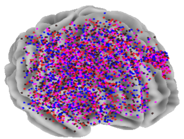
Firing HEC neurons plotted as coloured dots across a 3-D brain surface
Fig 4.9. Simultaneous firings of one million HEC neurons mapped on a brain surface. Each colour is one neurocomputational feature.
§4.5 · Spatial firing distribution

A million neurons, seen on the brain surface

A grid of one million HEC neurons starts at a resting potential of −80 mV, each with 1,000 sparse synaptic connections. Every neuron that reaches 30 mV is drawn as a dot at its (i, j, k) position, so the firing pattern can be read directly on the cortex.

Isynaptic = Σ 0.2 · rand(1, 1000)  (4.1)
106neurons
1,000synapses per neuron
5firing features

Homogeneous runs give all regions the same five features; heterogeneous runs give each region its own mix. The firings form propagating waves that appear and vanish across the surface, much as in Izhikevich's large-scale thalamocortical model. The same pipeline can be scaled up on SpiNNaker or the K computer and compared with EEG, MEG, ECoG and voltage-sensitive-dye recordings.

Beyond the brain · thesis §5.6

The same model in the pancreas

An extension from the thesis book: adding glucagon and insulin terms lets the same HEC equations describe the islet cells that control blood glucose.

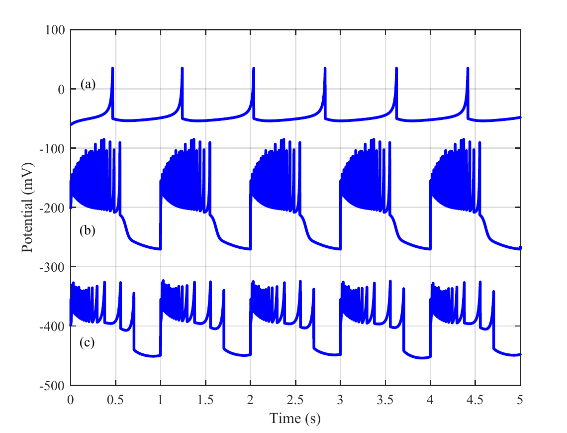
Simulated membrane potentials of pancreatic alpha, beta and delta cells over 5 seconds
Fig 5.30. (a) α-cells fire single spikes at low glucose. (b) β-cells and (c) δ-cells burst as insulin rises and decays. Traces are offset vertically.
§5.6 · Pancreatic cells

One model for glucagon and insulin secretion

Diabetes is driven by failures of glucose homeostasis, which the brain regulates through the autonomic nervous system. Adding glucagon- and insulin-induced potentials to the HEC equations reproduces the electrical activity of all three islet cell types.

α · glucagon, low glucoseβ · insulin, rising glucoseδ · insulin, falling glucose
CV′ = k(V−Vr)f(V−Vth) − u + Vα + Vβ + RI  (5.5)
u′ = a(bV − u)  (5.6)
Vth = Vglue−(t−nτ)/Tk + Vinse−(t−nτ)²/TCa  (5.8)
Model parameters (Table 5.3)
Parameterα-cellsβ-cellsδ-cells
a0.030.020.02
b−2−5−1
c050 μF50 μF
d1005060
k130.3
f0.10.050.2
Vglu035 mV35 mV
Vins030 mV40 mV
VCa070 mV70 mV
Rk05 MΩ5 MΩ
RCa05 MΩ5 MΩ
R95 MΩ95 MΩ95 MΩ

Integrated with the Euler method, V(k+1) = V(k) + hV′(k). Published in the Arabian Journal for Science and Engineering (2024).

From model to silicon · thesis book, chapter 6

NeuroBit64: the model as a family of chips

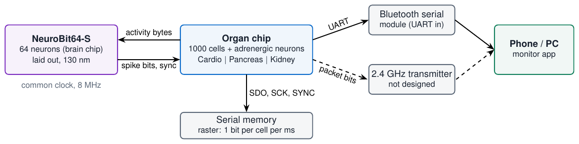
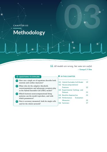
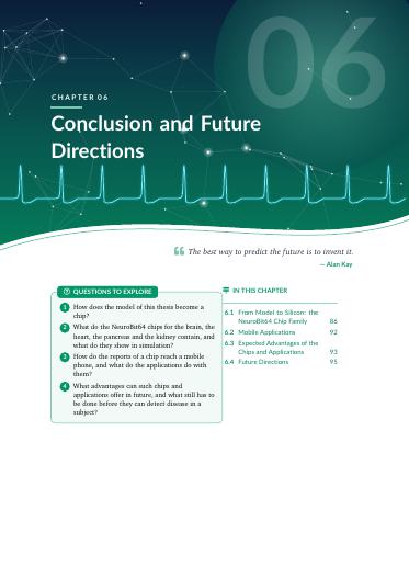
After the thesis, the models were written as digital circuits. One brain chip and three organ chips share one architecture, talk to each other over three wires and report to a phone over Bluetooth. All four are verified designs; none has been fabricated yet.

Live · the chips running in this page
Body Monitor: brain, heart, pancreas and kidney together

Press a button, or the Chips link in the menu, to start the chips here. They run the same integer models as the Verilog designs.

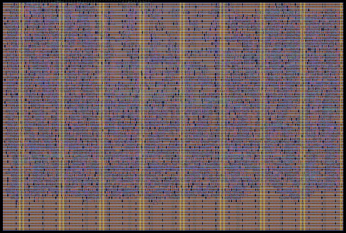
Mask layout of the NeuroBit64-S chip: rows of standard cells with metal routing
Layout of NeuroBit64-S. SkyWater 130 nm, about 0.33 × 0.22 mm, design-rule and layout-versus-schematic clean. It is the only chip of the family with a mask layout so far.
One architecture · four chips

A brain chip, organ chips and a phone

One arithmetic circuit serves all cells in turn, one cell per slot of 8 clocks. An organ chip holds 1000 cells; at 8 MHz one step of the whole network takes 1 ms, so it runs in real time.

  • Brain link: 32 group-activity bytes go to the 64 neurons of the brain chip; its 64 decision bits come back as sympathetic and parasympathetic drive.
  • Bluetooth: a 16-byte report every 16 ms for a Bluetooth serial module, and the bit stream of a Bluetooth LE advertising packet.
  • Verified: each chip's Verilog is identical, bit for bit, to a software model, also with the real brain chip in the loop.
Block diagram: NeuroBit64-S brain chip, organ chip, Bluetooth serial module, phone
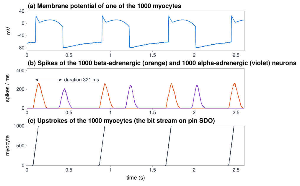
Three plots: membrane potential of one myocyte, population spikes of beta and alpha neurons, and a raster of 1000 myocytes over 2.6 seconds
NeuroBit64-Cardio, normal rhythm. β neurons fire at the upstroke, α neurons at the end of the plateau; the time between the two bursts is the action potential duration.
Brain and heart

Rhythm disorders, read from adrenergic neurons

The chip measures beat interval, beat-to-beat change and action potential duration from the neuron populations alone, and raises seven rhythm flags.

79 beats/min, normal35 ms RMSSD314 ms action potential

In built-in test conditions it flags tachycardia (137 beats/min), bradycardia (50), an irregular rhythm, a conduction block and a prolonged action potential. With the brain chip connected, the returned drive changes the heart rate: the loop is closed.

These are conditions of the model, not tests on patient recordings.

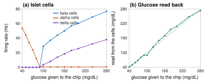
Two plots: firing rate of beta, alpha and delta cells against glucose, and glucose read from the cells against glucose given
NeuroBit64-Pancreas, healthy islet. α-cells are active at low glucose, β- and δ-cells at high glucose; the chip reads the glucose level back from them.
Brain and pancreas

The glucose level, read from the islet

Given a glucose value from a sensor, the network of islet cells produces its own insulin and glucagon levels. The glucose read back from the cells differs from the value given by 8 mg/dL on average (40–295 mg/dL).

When β-cells are lost or respond weakly, or α-cells do not respond, the read-back no longer follows the glucose, and the chip flags it.

The glucose value comes from a sensor: the companion project GlucoSense is a wearable glucose-monitor application with its own sensor-chip design.

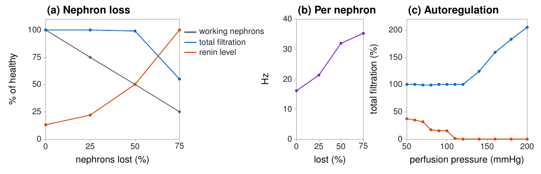
Three plots: working nephrons, total filtration and renin against nephron loss; rate per nephron; filtration against perfusion pressure
NeuroBit64-Kidney. With 25 % or 50 % of the nephrons switched off, total filtration stays normal because the remaining ones work harder.
Brain and kidney · exploratory

The early stage that total filtration hides

The chip counts the working nephrons and the load on each. Losing 25 % or 50 % of them leaves total filtration at 99–100 %, but the chip raises “nephrons missing” and “hyperfiltration” together: an early-loss pattern.

This model is new. It is not part of the thesis and has not been compared with kidney data; whether such a pattern can be measured in a real kidney is an open question.

Mobile applications

From the chip to the phone

A chip that listens to an organ is useful only if its report reaches the person who needs it. The phone is the natural display: it already has a Bluetooth radio and a screen, and the applications are web pages that need no installation.

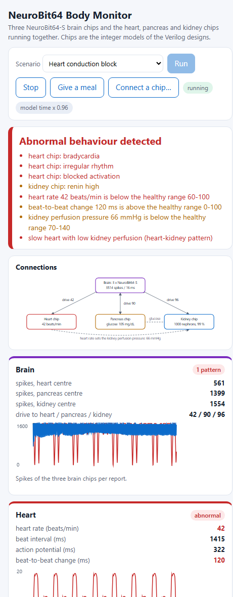
Phone screenshot of the Body Monitor reporting abnormal behaviour with a list of findings
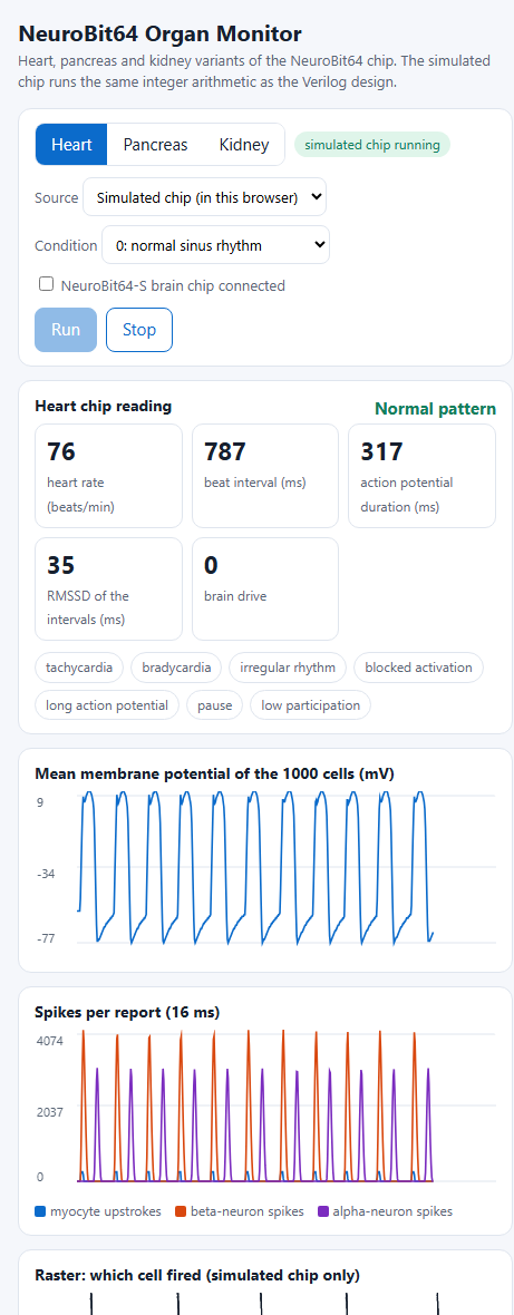
Phone screenshot of the Organ Monitor showing heart rate, beat interval and action potential duration
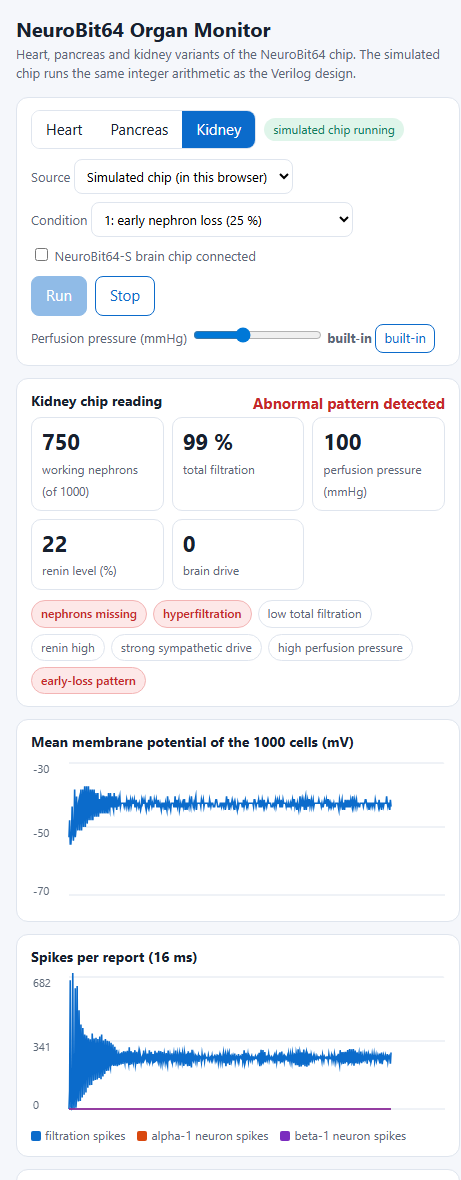
Phone screenshot of the Organ Monitor showing 750 working nephrons and the early-loss flags
Three applications

What the applications do

  • The path: every 16 ms the chip writes a 19-byte frame on a serial pin; a small Bluetooth serial module forwards it; the application checks the checksum and decodes the report. 62 reports per second are only about 1.2 kB/s.
  • Organ Monitor: one chip at a time. Heart: rate, beat interval, action potential duration, RMSSD. Pancreas: glucose given, glucose read from the cells, insulin level. Kidney: working nephrons, filtration, pressure, renin. The flags are listed by name.
  • Body Monitor: three brain chips with heart, pancreas and kidney chips together. An anomaly detector combines the chips' flags, nine values against their healthy range and four patterns across organs, and reports normal, watch or abnormal with the findings in plain words.
  • GlucoSense: the companion glucose-sensor application; with the pancreas chip it forms a chain from sensor to islet model to phone.

The applications also run the chips' own models, bit-identical to the Verilog, so they work before any hardware exists. That is how they have been tested; the Bluetooth path has not been tried with a real chip.

Looking ahead

What such chips and applications can offer

These are expectations that follow from the designs. Each has to be demonstrated with fabricated chips and with data from subjects.

Continuous, in real timeA clinic visit is a short sample of a long process. A chip that steps the organ model every millisecond follows the organ without interruption and reports every 16 ms. Early detectionThe models watch relations between signals, not one number. In simulation, losing half of the nephrons left total filtration normal, yet the chip saw it. Small, low power, low costPurely digital circuits of a few mm² in a mature 130 nm process at 8 MHz: 1000 cells share one arithmetic circuit. That is the condition for a wearable patch and, later, an implant. No special equipmentThe display is a phone and the applications are web pages: usable by a basic health unit, a family doctor or the patient at home, where laboratories are far away. ExplainableEvery flag has a definition, such as “beat interval above 1000 ms” or “filtration per working nephron above 125 %”. A physician can check why it was raised. PrivateSignals are processed on the chip and shown on the user's own phone. Nothing has to go to a server for the result to appear. The whole bodyHeart, pancreas and kidney influence each other. Chips with a common brain link and report format can be combined, so disorders can be seen in the relation between organs. One architecture, many organsA further organ needs a cell model and parameters, not a new chip: link, report, Bluetooth and applications are reused. Open and reproducibleDesigns, models, tests and applications are public, and the brain chip's layout was made with free tools. Others can repeat, correct and extend the work.

The same architecture is planned for further organs (lung, gut, liver, muscle, retina). Next steps: fabrication, and validation of each model against recorded data.

Publications

Five papers, what each one did

First author on all five. Use Copy citation to paste a reference.

PhD thesis · book edition

Read the whole thesis as a book

The complete thesis, redesigned for reading: a full-colour layout, opening questions and key takeaways for every chapter, and an appendix with the MATLAB code behind each figure.

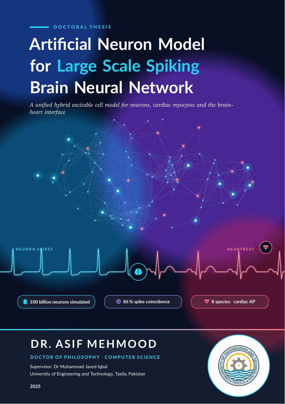
Front cover of the thesis book: Artificial Neuron Model for Large Scale Spiking Brain Neural Network, by Dr. Asif Mehmood

Artificial Neuron Model for Large Scale Spiking Brain Neural Network

Dr. Asif Mehmood · PhD Computer Science · UET Taxila · 2025, chapter 6 extended 2026 · 139 pages · PDF 5.7 MB

One hybrid excitable cell model, followed from ion channels to a network of 100 billion neurons. It covers neurons, cardiac myocytes of eight species and pancreatic cells, a cardio-neural network that predicts heart rate variability, and, in the extended chapter 6, the NeuroBit64 family of chips for brain, heart, pancreas and kidney.

  1. 1Introduction
  2. 2Literature Review
  3. 3Methodology
  4. 4Results: Neuron
  5. 5Results: Cardiac Myocyte
  6. 6Conclusion and Future Directions
  7. AMATLAB Source Code
From the thesis

Figures

Artificial Neuron Model for Large-Scale Spiking Brain Neural Network, PhD thesis, UET Taxila, 2025. Select a figure to enlarge it.

Curriculum vitae

Education and experience

  1. 2017–2025

    PhD in Computer Science, UET Taxila

    Thesis: Artificial Neuron Model for Large-Scale Spiking Brain Neural Network. Approved July 2025, conferred September 2025. Supervisor: Dr Muhammad Javed Iqbal.

  2. 2011–2014

    MS Computational Science & Engineering, NUST

    Research Centre for Modeling and Simulation, Islamabad. CGPA 3.55/4.00. Thesis: Electrical Modeling of Excitable Cells (grade A): neurons and cardiac myocytes as equivalent electronic circuits.

  3. 2009–now

    Lecturer, Electrical & Electronics Engineering

    COMWAVE Institute of Sciences and IT, Haripur (Approved Education Centre of SUIT Peshawar). Bachelor-level courses, final-year project supervision, curricula and lab manuals under OBE.

  4. 2017

    B.Ed., Virtual University of Pakistan

  5. 2006–2008

    MSc Electronics, Quaid-i-Azam University

    Islamabad.

  6. 2003–2005

    BSc Mathematics, Physics, Electronics, Hazara University

    Mansehra. First Division.

Research interests

Brain & body modelling
Computational & systems neuroscienceBrain–heart–pancreas interactionCardiac & metabolic electrophysiologyPhysiological digital twinsBrain-scale simulation & HPC
Intelligent systems
Neuromorphic computing & spiking neural networksExplainable AI for healthcareAnomaly & early-disease detectionEdge AI / TinyML
Biomedical technology
Biosensor integration (EEG, ECG, glucose)Sensor interfacing & embedded systemsBiomedical & electronic signal processingWearable & implantable devicesBrain–computer interfacesAndroid mHealth apps & remote monitoring

Technical skills

Programming
Python, MATLAB, C/C++
Modeling
Spiking and conductance-based neuron models, large-scale network simulation, machine learning, anomaly detection
Signals
EEG and ECG analysis, electrophysiological recordings
HPC
OpenMP; CUDA (introductory)
Hardware
Verilog/VHDL, ModelSim, SPICE; UPPAAL and PRISM model checking
Writing
LaTeX

Training

IBRO
Four months: From Basic Neuroscience to Advanced AI Applications, with HPC
NUST
One month: programming supercomputers for AI (OpenMP, CUDA)
NUST
Six weeks: machine learning for large-scale system analysis in MATLAB
Contact

Open to postdoctoral and faculty positions

Computational neuroscience, spiking or neuromorphic AI, and biomedical modeling. Referee details are available on request.

Emailasifnazquaidian@gmail.com
ORCID0000-0003-4924-9854
Open ↗
Based inHaripur, Pakistan